Home
/ Plant Cell Electron Microscope Images - Electron Tomography Of Cryo Immobilized Plant Tissue A Novel Approach To Studying 3d Macromolecular Architecture Of Mature Plant Cell Walls In Situ : A scanning electron microscope (sem) is a type of electron microscope that produces images of a sample by scanning the surface with a focused beam of electrons.
Plant Cell Electron Microscope Images - Electron Tomography Of Cryo Immobilized Plant Tissue A Novel Approach To Studying 3d Macromolecular Architecture Of Mature Plant Cell Walls In Situ : A scanning electron microscope (sem) is a type of electron microscope that produces images of a sample by scanning the surface with a focused beam of electrons.
Plant Cell Electron Microscope Images - Electron Tomography Of Cryo Immobilized Plant Tissue A Novel Approach To Studying 3d Macromolecular Architecture Of Mature Plant Cell Walls In Situ : A scanning electron microscope (sem) is a type of electron microscope that produces images of a sample by scanning the surface with a focused beam of electrons.. Cells of plant or animal tissue. However, light microscopes form real colour images and can be used to watch living processes occur in microscopic detail, while electron microscopes cannot be used to study living cells. The left image is obtained from the electron microscope and it contains few bright dots and few dark dots as image contrast. It is a special type of microscope having a high resolution of images, able to magnify it provides detailed images of the surfaces of cells and whole organisms that are not possible by tem. Wells, plates, covers, tissue culture inserts.
Collection by chris • last updated 3 weeks ago. Light and electron microscopes allow us to see inside cells. Cancer cells (hela), imaged by electron microscope. However, light microscopes form real colour images and can be used to watch living processes occur in microscopic detail, while electron microscopes cannot be used to study living cells. Cells of plant or animal tissue.
Plant Cell Under Electron Microscope Diagram Quizlet from o.quizlet.com See more ideas about microscopic photography, electron microscope, electron microscope images. The protrusion at center is just over 50 microns tall. Cells of plant or animal tissue. Chemicals for electron microscopy, light microscopy and histology. It is very difficult to find which dot belongs to which atom. Level suitable for as biology. The latest media tweets from unseen world (@micropicx). Science nature micro photography cell image things under a microscope image of the day microbiology microscopic.
It is very difficult to find which dot belongs to which atom.
Category:electron microscope images (en) categoría de wikimedia (es); For images showing electron microscopes see category:electron microscopes. Cells of plant or animal tissue. The left image is obtained from the electron microscope and it contains few bright dots and few dark dots as image contrast. Amazing images through the eyes of the electron microscope. It is very difficult to find which dot belongs to which atom. Microscopic genetics microscope biology notes science plant science mitosis cell biology cell. However, light microscopes form real colour images and can be used to watch living processes occur in microscopic detail, while electron microscopes cannot be used to study living cells. Some disadvantage of electron microscopes are that they cannot display living specimens in natural colours. Browse 441 electron microscope cells stock photos and images available, or start a new search to explore more stock photos and images. The colours are added afterwards using specialised computer software. All of the organelles in plant and animal cells can be seen under a light microscope. See more ideas about electron microscope images, electron microscope, microscopic photography.
Electron microscope cell stock photos and images. A scanning electron microscope (sem) is a type of electron microscope that produces images of a sample by scanning the surface with a focused beam of electrons. Cells of plant or animal tissue. See more ideas about microscopic photography, electron microscope, electron microscope images. An electron microscope is a type of microscope that uses electrons to illuminate a specimen and create an enlarged image.
Plant Cell Tem Stock Image C038 7324 Science Photo Library from media.sciencephoto.com Science nature micro photography cell image things under a microscope image of the day microbiology microscopic. The final image produced from an electron microscope is always in greyscale; Find the perfect electron microscope cell stock photo. A scanning electron microscope (sem) is a type of electron microscope that produces images of a sample by scanning the surface with a focused beam of electrons. Plant cells and tissues scanning electron microscope image of a leaf from a black walnut tree. Plant stem plant cell plant science science art biology art microscopic photography microscopic images cross section abstract watercolor art. This is the ability to see two points as two points, rather. See more ideas about microscopic photography, electron microscope, electron microscope images.
Electron microscopes have much greater resolving power than light microscopes and can obtain much higher magnifications.
In this microscope, images are produced from the interaction between the prepared samples in the vacuum. Category:electron microscope images (en) categoría de wikimedia (es); Microscopic genetics microscope biology notes science plant science mitosis cell biology cell. The protrusion at center is just over 50 microns tall. An electron microscope is a type of microscope that uses electrons to illuminate a specimen and create an enlarged image. Wood cells of holly (source). An electron microscope is a microscope that uses a beam of accelerated electrons as a source of illumination. See more ideas about electron microscope, electron microscope images, microscopic images. Level suitable for as biology. Plant cells and tissues scanning electron microscope image of a leaf from a black walnut tree. Electron microscopes use electron beams focused by electromagnets to magnify and resolve microscopic specimens. The scanning electron microscope (sem) has seldom been used to generate images for the purposes of analysis, largely because conventional imaging of biological. Cells of plant or animal tissue.
Below you will find a small collection of images from scientists around the. See more ideas about microscopic photography, electron microscope, electron microscope images. Find the perfect electron microscope cells stock photos and editorial news pictures from getty images. See more ideas about electron microscope, electron microscope images, microscopic images. Light and electron microscopes allow us to see inside cells.
Galleries Biological Sciences from www.ualberta.ca See more ideas about electron microscope images, electron microscope, microscopic photography. Find the perfect electron microscope cell stock photo. Electron microscopes have much greater resolving power than light microscopes and can obtain much higher magnifications. An image of a single cell of the plant pathogenic bacterium, pseudomonas syringae, is presented in fig. Wood cells of holly (source). Cells of plant or animal tissue. Cancer cells (hela), imaged by electron microscope. In this microscope, images are produced from the interaction between the prepared samples in the vacuum.
Light and electron microscopes allow us to see inside cells.
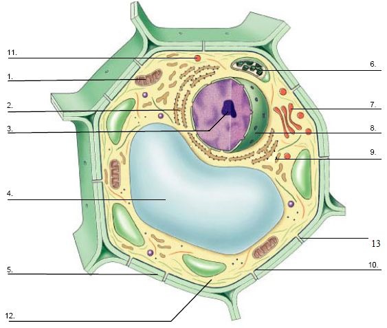
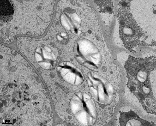
It is a special type of microscope having a high resolution of images, able to magnify it provides detailed images of the surfaces of cells and whole organisms that are not possible by tem. When you look at animal or plant cells under the electron microscope, you can see a lot more detail. Plant cells and tissues scanning electron microscope image of a leaf from a black walnut tree. Science nature micro photography cell image things under a microscope image of the day microbiology microscopic. Transmission electron microscope image of a chloroplast. Complete with scanning electron microscope images. Level suitable for as biology. Wells, plates, covers, tissue culture inserts. An image of a single cell of the plant pathogenic bacterium, pseudomonas syringae, is presented in fig. On plant leaves, there is a strong selection for cells with traits that would also favour survival in the atmosphere, such as coping with conditions of variable water availability, extensive radiation or. Below you will find a small collection of images from scientists around the. An electron microscope is a microscope that uses a beam of accelerated electrons as a source of illumination. See more ideas about electron microscope, electron microscope images, microscopic images.
An electron microscope is a microscope that uses a beam of accelerated electrons as a source of illumination plant cell microscope image. It is very difficult to find which dot belongs to which atom.
Share :
Post a Comment
for "Plant Cell Electron Microscope Images - Electron Tomography Of Cryo Immobilized Plant Tissue A Novel Approach To Studying 3d Macromolecular Architecture Of Mature Plant Cell Walls In Situ : A scanning electron microscope (sem) is a type of electron microscope that produces images of a sample by scanning the surface with a focused beam of electrons."


Post a Comment for "Plant Cell Electron Microscope Images - Electron Tomography Of Cryo Immobilized Plant Tissue A Novel Approach To Studying 3d Macromolecular Architecture Of Mature Plant Cell Walls In Situ : A scanning electron microscope (sem) is a type of electron microscope that produces images of a sample by scanning the surface with a focused beam of electrons."